Cardiac Catheterization Imaging
At St. Peter’s Health Partners, you will find advanced heart services close to home, including imaging tests performed inside the heart.
Why Choose St. Peter’s Health Partners for Your Cardiac Catheterization Imaging?
Cardiac catheterization is a procedure that uses long tubes (catheters) to access the heart through small incisions. Tiny instruments at the catheter tip allow us to perform sophisticated evaluations that are not possible using other means. This information enables us to deliver high-quality care that helps more people achieve outstanding results.
Our team includes cardiologists with additional training in catheter-based techniques, including imaging.
This level of care is not widely available
in the Capital Region.
Looking For Test Results?
View test results once they are uploaded by your doctor.


Programs with a high volume of heart catheterizations have higher success rates and ours is no exception. We perform approximately 5,000 interventional cardiology procedures per year. This level of experience leads to precise assessments and reliable results.
Interventional cardiologists are part of our talented cardiology team. They work alongside radiologists, heart surgeons, and other specialists to coordinate your care. Our team approach helps you receive services that are appropriate for your circumstances.
Types of Cardiac Catheterization Imaging
Types of Imaging We May Use During Cardiac Catheterization Procedures Include:
- Fractional Flow Reserve (FFR) measures the blood pressure and flow through the coronary arteries. We use it to pinpoint minor blockages and assess their impact on oxygen flow.
- Intracardiac Echocardiography provides real-time imaging of heart structures using sound waves. A tiny catheter with an ultrasound sensor is passed into the heart to assess atrial fibrillation (AFib) and other arrhythmias and for treating heart valve disease.
- Intravascular Ultrasound (IVUS) provides enhanced views of coronary arteries using high-frequency sound waves. We use IVUS to measure the degree of blood vessel narrowing in people with coronary artery disease.
- CT Angiography combines a CT scan with a special dye that enhances views of your blood vessels. A CT scan takes images from multiple angles and a special computer combines them. This type of imaging test helps us identify an aortic aneurysm, or an abnormal bulge in a blood vessel wall.
- Optical Coherence Tomography (OCT) uses infrared light to view blood vessels and provides a significantly more precise picture than an intravascular ultrasound. We sometimes use it to guide therapies, such as stent placement, to keep narrowed arteries open.
Cardiac Catheterization Imaging: What to Expect
Here’s What Happens During Cardiac Catheterization Procedures:
- We give you medication to help you relax. Some people fall asleep. Even if you stay awake, you will be comfortable and experience no discomfort.
- An interventional cardiologist makes a small incision. Depending on the type of imaging you need, it may be in your groin, wrist, or neck.
- We place a catheter through the incision and advance it to reach the heart.
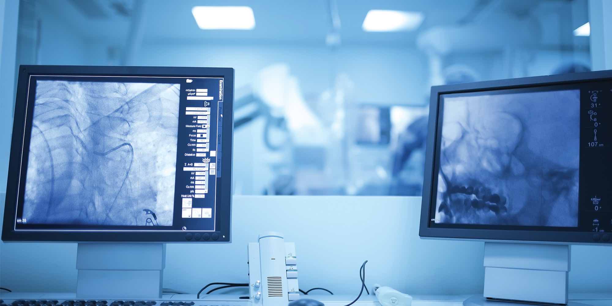
- Real-time imaging shows the precise location of the catheter on a large screen.
- Our experts conduct the imaging test you need with magnified results showing on a separate high-definition screen.
- If tests involve injecting a dye through a blood vessel, you may experience sudden warmth or flushing as the dye enters your bloodstream.
- When the procedure is complete, we remove the catheter. A bandage may be all you need to protect the incision while it heals.
Take the Next Step
Call 1-800-HEART-76 (1-800-432-7876) to find a heart specialist or learn more about cardiovascular care at St. Peter’s Health Partners.