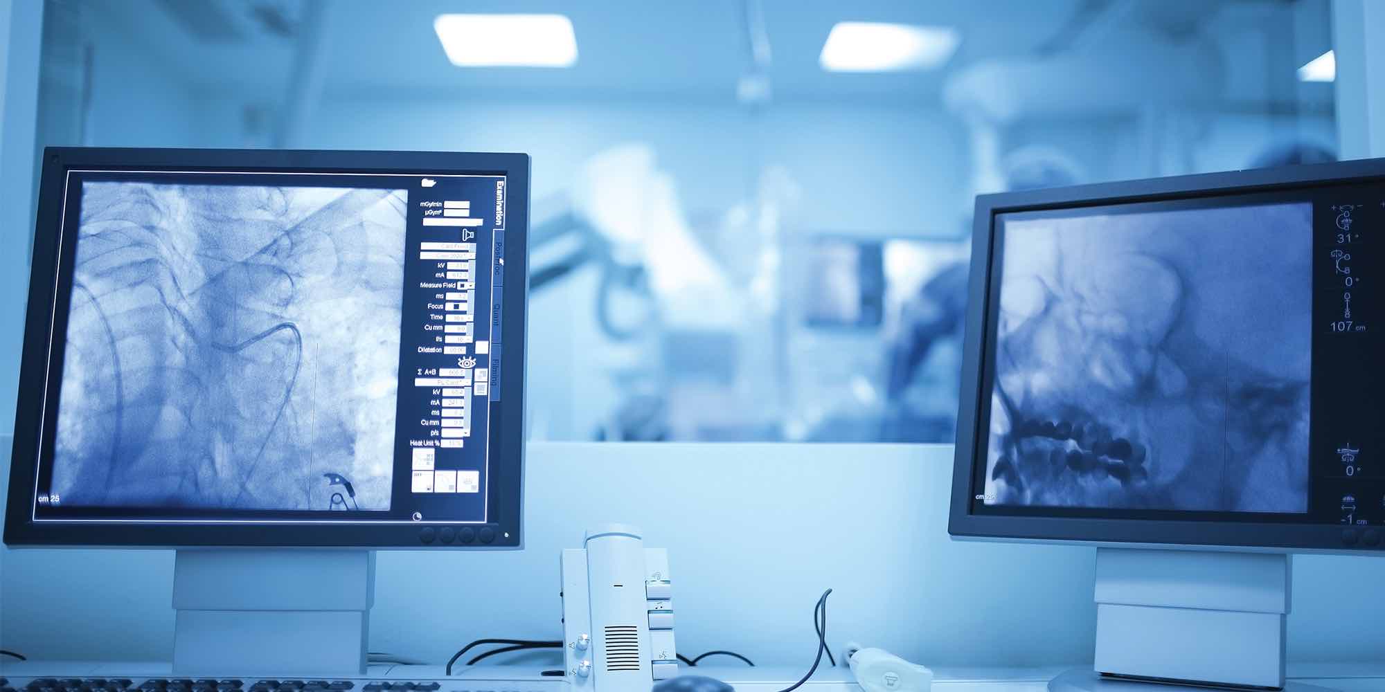
Cardiac Imaging
Cardiac imaging is one of the many ways St. Peter’s Health Partners cares for people with heart disease symptoms. We offer convenient access to a broad range of options.
Why Choose St. Peter’s Health Partners for Cardiac Imaging?
Our team includes doctors specializing in abnormal heart rhythms, valve disease, heart failure, and more. We use research-based methods to determine the best option for assessing your symptoms. Our team explains how tests work, why we are recommending them, and what to expect. Find out more about the heart conditions we treat.
We hold ourselves to the highest heart imaging standards, which ensures you receive safe, precise testing. This level of care enables us to maintain accreditation from the Intersocietal Accreditation Commission (IAC).
Heart Imaging Procedures
We also offer minimally invasive heart imaging procedures. These tests use small incisions and sophisticated technologies to assess heart structures and function. You may need one of these tests if noninvasive imaging studies identify an issue and we need more information. Find out more about cardiac catheterization imaging.
Looking For Test Results?
View test results once they are uploaded by your doctor.
Heart Imaging Available at St. Peter’s Health Partners
St. Peter's offers cardiac imaging studies at our acute care facilities as well as our medical offices and clinics. These include:
- Chest X-ray use radiation to capture images of the bones, tissue, and blood vessels in your chest. We use it to evaluate the heart and lungs for abnormal structures, fluid buildup, and more.
- Heart Computed Tomography (CT) Scan takes X-rays from different angles. A computer assembles the images to create sophisticated views of the heart’s tissue and blood vessels.
- Stress Echo uses sound waves to evaluate function when the heart is working harder than usual. During the test, you walk at a gentle pace on a treadmill. If you are too sick to go on a treadmill, we give you medication to simulate the effect of exercise on your heart.
- Echocardiography (Echo) uses high-frequency sound waves to assess the size and thickness of heart tissue and whether there is extra fluid in the heart. We also use the test to evaluate the heart’s pumping function and force of blood through the blood vessels. This test is also known as a transthoracic echocardiogram.
- Transesophageal Echo (TEE) During TEE, we slide a narrow probe down your throat and into the tube that connects it to the stomach (esophagus). This method helps us capture clearer images than a standard echo because the esophagus is near the heart. We use TEE to detect signs of infection, assess whether blood is flowing in the proper direction, and more.
Take the Next Step
Call 1-800-HEART-76 (1-800-432-7876) to find a heart specialist or learn more about cardiovascular care at St. Peter’s Health Partners.


